
Cardiovascular Disease
In 2019, the American Heart Association reported that at least 48 percent of U.S. adults have some form of cardiovascular disease, and many times there are no clear warning signs. The stereotype scene of clutching your chest and falling to the floor does not always happen. Some symptoms do not even occur in your chest!
But there are everyday signs which can identify possible blockage. These include:
- Nausea, Indigestion, Heartburn, or Stomach Pain
- Dizziness or Lightheadedness
- Easily exhausted
- Breaking out in a cold sweat
- Swollen legs, feet, and ankles
- Pain between the shoulder blades
Experiencing any of these symptoms does not automatically mean you have a heart condition. But, if you are not sure, it is essential to talk with your primary care provider. This is especially important if you are 60 years or older, overweight, or have diabetes, high cholesterol, or high blood pressure.
After visiting your healthcare provider and having a few routine tests run, you may be referred to a cardiologist. This heart expert will start with a physical exam, including blood work to measure cholesterol and blood sugar levels. Depending on your health history, additional tests may be ordered.
Exercise Stress Test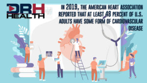
A stress test will show how well your heart handles work. As your body works harder during the test, your heart must pump more blood as you need more oxygen. The test can show a reduction in blood supply in the arteries that supply the heart. It also helps to show the kind and level of exercise appropriate for you.
Holter Monitor
If your symptoms suggest that an occasionally irregular heart rhythm may be causing your condition, it may be recommended that you wear a Holter monitor for a day or so. This monitor captures and displays the heart’s performance and often detects abnormal heart rhythm (arrhythmias).
Echocardiogram
An echocardiogram uses sound waves to produce images of your heart. This standard test provides detailed information on your heart size, shape, heart valves working correctly, and if your heart valves are too narrow.
Transesophageal Echocardiogram (TEE)
Transesophageal echocardiography (TEE) is a test that produces much more detailed pictures of your heart. TEE uses high-frequency sound waves (ultrasound) to make detailed pictures of your heart and the arteries that lead to and from it. TEE can give more explicit images of the heart’s upper chambers and the valves between the heart’s upper and lower chambers than standard echocardiograms.
If determined that you have significant narrowing of the arteries, there are several treatment options available. Some are treated with medicine or with a non-surgical option, a stent. Some people may require bypass surgery, depending on the number of blockages, the location, and the complexity.
Cardiac Stent
A stent is a tiny wire mesh tube. It props open an artery and is left there permanently.
A needle is inserted with a catheter that guides specialized tools through your blood vessels to reach the area requiring a stent. This incision is typically in the groin or arm. Once the broken or blocked vessel is located, the stent is put into place.
Additional procedures are available to treat heart conditions, including Catheter Ablation and Maze Procedure.
Many patients only consider seeing a doctor when they don’t feel well, or they may wait for an annual physical with their family care doctor to ask questions about any physical pains or symptoms. But if you have concerns about your heart, please contact your healthcare provider to discuss these issues.
Kris Mullins, MD
Interventional Cardiology
Pavilion Clinic
580.251.6806